Hypothesis
A Genome the Thymus Never Read
Private mitochondrial mutations and the geography of autoimmune disease.
Somewhere in your body right now there is very likely a T cell that recognizes a fragment of your own mitochondria and would attack the cell displaying it, if the circumstances lined up. That is not a thought experiment. In 2014 a group at York University in Toronto drew blood from healthy volunteers, people with no autoimmune diagnosis of any kind, and found circulating T cells that recognized and responded to peptides generated by mutated mitochondrial DNA. The cells were already present, and they were already primed. The donors were fine. What follows is an argument about the difference between those healthy donors and the patients who fill a rheumatology clinic, and about whether that difference is partly a question of which mutations landed in which tissue, and when.
Let me hold that finding for a moment and lay out the material it stands on.
A genome that keeps changing
Every cell carries mitochondria, and every mitochondrion carries its own small circular genome: thirty-seven genes, thirteen of which code for protein subunits of the machinery that makes ATP. This genome is not a museum piece. It mutates, and because a single cell holds hundreds to thousands of copies, mutant and normal versions coexist in the same cell, a state called heteroplasmy. Over a lifetime the mutant fraction in a given tissue can climb, sometimes because a mutated genome gains a slight replicative advantage and its descendants take over a cell or a patch of neighboring cells. The pattern is not uniform across the body. Deep sequencing of multiple organs from the same person shows that kidney, liver and skeletal muscle accumulate characteristic variants that are present in one tissue and undetectable in another from the same individual. Your liver at sixty is, in a small but real sense, running slightly different software than the liver you were born with, and different software than your kidney.
The lesson the thymus teaches
Central tolerance is the reason you do not, under ordinary conditions, attack yourself. Early in life, developing T cells are marched through the thymus and shown a broad sample of the body’s own proteins. Any T cell whose receptor binds a self-peptide too avidly is deleted, or converted into a regulatory cell. The catch, and it is the hinge of this whole argument, is that the thymus can only tolerize against peptides it actually presents. This is not a new idea in the abstract. In type 1 diabetes, neoepitopes created by chemical modification of beta-cell proteins escape tolerance precisely because the modified forms never appear in the thymus. A mouse model of autoimmune hepatitis makes the same point with a liver peptide: absent from the thymus, it fails to delete its matching T cells, which then accumulate in the periphery.
Now set the mitochondrial genome inside that frame. The thymus presents peptides derived from whatever mitochondrial sequences its own cells happen to carry. It cannot present a peptide from a missense mutation that will arise decades later in a distant organ, because that sequence did not exist when the lesson was taught. A T cell capable of recognizing that future variant is never removed. The variant is private, belonging to a single person’s organ at a particular age.
The thymus tolerized you against whatever mitochondria it happened to contain. What your kidney would be carrying at sixty was never on the syllabus.
I will give this object a name, because the obvious names are already taken. “Mitochondrial neoantigen” refers, in the lupus literature, to mitochondrial RNA and DNA acting as antigens; “neoself antigen” refers to a separate misfolding phenomenon. Call the thing at issue here a private somatic mitochondrial neoepitope: a peptide from a clonally expanded missense mutation in one of the thirteen protein-coding mitochondrial genes, present in a peripheral tissue and absent from the thymic repertoire.
Is presentation of such peptides even plausible? Mouse immunology answers cleanly. There is a dedicated molecule, H2-M3, that specializes in presenting short formylated peptides taken from the very N-termini of the thirteen mitochondrially encoded proteins. It discriminates between variants differing by a single residue, and the T cells it educates are selected in the thymus according to which mitochondrial peptides are on display there. The human situation is messier. H2-M3 has no clean human counterpart, and the human evidence rests instead on the classical HLA molecules used in the Toronto study. Still, the principle that a single-residue mitochondrial variant can become a T-cell target is not speculation. It has a crystal structure.
Why mitochondria, of all things
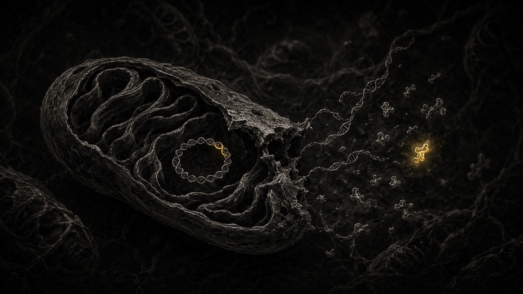
An autoreactive T cell needs two things to start a fire: an antigen to aim at, and a danger signal telling the immune system this is not routine housekeeping. In most models these two arrive separately and have to coincide. Mitochondria supply both from a single source. When one ruptures, in necrosis or in the neutrophil death called NETosis, it releases its DNA, and oxidized mitochondrial DNA is a potent trigger of the cGAS-STING and TLR9 pathways that drive type I interferon, the signature seen across systemic autoimmune disease. The same rupture releases the mutant peptide. Adjuvant and antigen come out of one broken organelle, in the same place, at the same instant. Cancer vaccinologists have already turned this to use, engineering antigens into mitochondria because the organelle self-adjuvants once a phagocyte swallows it. The proposal here runs the logic backward, toward a private antigen nobody engineered, arising in a person’s own kidney.
What the idea would explain
Three features of systemic autoimmune disease sit awkwardly in current models, and this one reaches all three without extra machinery.
The first is organ tropism. If the driver is a variant that clonally expanded in a particular tissue, the disease should localize where the clone did.
The second is age. Heteroplasmy accumulates over decades, so later onset follows from the biology of the substrate. There is a sharper reason as well. The thymus involutes; by middle age its output is a fraction of what it was, and the mitochondrial repertoire it fixed as “self” belongs to a body that no longer exists in molecular detail. Tolerance was calibrated against a snapshot, and the tissues kept moving after the shutter closed. The mismatch between the frozen reference and the drifting periphery widens with every passing year. That is a more specific claim than the familiar observation that an aging thymus simply tolerizes less well. It does not even require the thymus to fail, only that the periphery keeps changing after the lesson is over.
The third is the maternal skew. Mitochondria come from the mother, and a large Taiwanese population study found that children of mothers with autoimmune disease carry an elevated risk of lupus in particular. This is the weakest of the three legs, and it deserves to be marked as such. Maternal inheritance is confounded by the X chromosome and by the intrauterine environment. Microchimerism muddies it further. The same “maternal bias implies mitochondria” inference was tested directly in familial Parkinson’s disease and failed there. I offer the maternal skew as consistent with the model, not as evidence for it.
The prediction that decides it
Here is where the idea earns or loses its keep. For years, investigators have hunted for associations between inherited mitochondrial haplogroups and diseases such as lupus and rheumatoid arthritis. The results have been thin and hard to replicate, the kind of literature that gets waved off as underpowered noise. If autoimmune risk of this sort were carried by a germline haplogroup, that failure would be a disappointment. But if the true driver is a private, somatic variant that arose in one person’s tissue and appears in no one else’s germline, then a study built on germline haplogroups is looking in the wrong place by construction, and it has to come up empty. The weak haplogroup literature stops being an embarrassment and becomes a positive prediction.
A negative result that everyone files under noise is the result this model predicts.
A target that moves
Classical epitope spreading describes an immune response broadening over time to recognize more and more self-antigens. That spreading happens on the immune side. The model here adds a second kind of drift, on the side of the antigen itself. As a clone expands and further mutations accumulate, the tissue’s antigenic repertoire is rewritten while the disease is already underway. The closest precedent is not in autoimmunity at all but in oncology, where tumors evolve their neoantigen landscape under immune pressure. Carry that idea into non-malignant tissue and it yields a longitudinal prediction that can be checked: new variants in the affected organ should appear before new T-cell specificities, not after. A human template for the tracking already exists. In autoimmune lymphoproliferative syndrome, a somatic mutation in the FAS gene can be followed over years, its variant fraction rising and falling in lockstep with disease activity.
How to prove it wrong
A hypothesis you cannot kill is not worth much, so here is how to kill this one. Sequence mitochondrial DNA at single-cell or single-molecule resolution in affected tissue and in blood from the same patient, and, where tissue can be obtained, in the thymus. The model predicts a higher burden of missense variants in the coding genes in the target organ than in blood. Then look for T cells reactive to the specific mutant mitochondrial peptides and ask whether they are enriched in patients relative to controls. The decisive test is whether the expanded variant is private. If patients turn out to share the same expanded variant, the private-somatic story collapses, and what remains is an ordinary germline haplogroup effect wearing a disguise.
Where it could break
Several things could sink this, and they belong on the table rather than buried in a footnote. The cleanest mechanistic evidence, H2-M3, is a mouse molecule without a tidy human equivalent. Presentation of mitochondrial peptides on classical human HLA is quantitatively modest and depends on which HLA alleles a person carries, which would make the model’s penetrance ragged and patchy, though that raggedness matches how partial and incomplete real autoimmune risk actually looks. There is a direct hit to the word “private,” too. Some tissue-specific mitochondrial variants turn out to be recurrent across unrelated people, and those recurrent ones cluster in the non-coding regions that regulate replication rather than in the coding genes. The model has to be confined to private missense changes in the thirteen coding genes and kept well clear of that recurrent regulatory class. One further complication: perturbing mitochondria inside a regulatory T cell does not always weaken it. In at least one careful study, mitochondrial DNA release inside a regulatory T cell increased its suppressive output. The consequences of a damaged mitochondrion are not uniform, and any honest version of this model has to live with that.
If it holds
Suppose some version survives contact with the data. The therapeutic implication is an unusual one, because it points away from blunting the immune system and toward reducing the supply of antigen. The tools already exist, borrowed from other fields. Compounds that enhance mitophagy, such as urolithin A and precursors of NAD+, clear damaged mitochondria and are already in human use for aging muscle and for neurodegeneration. Techniques that shift heteroplasmy back toward the normal sequence, built originally for classic mitochondrial disease, can discriminate variants down to a single base pair. None of these was designed to treat autoimmunity. The reframing is what would be new: lower the private-neoepitope burden in a tissue before a clone expands past the point where enough peptide is presented to matter. A tissue’s mitochondrial mutational load would become something worth measuring.
The whole argument rests on one unproven step: that a private missense mutation, clonally expanded in a single organ, can generate a peptide the thymus never taught the immune system to ignore. Everything around it is already in the literature. The mitochondrial DNA acting as a danger signal, the tissue-specific accumulation of variants, the healthy donors carrying primed T cells against their own mutant mitochondria, all of it sits in separate papers that have not yet been asked to speak to one another. The experiment that would connect them is not exotic. It asks for affected tissue and a matched blood sample from the same patient, read on a sequencer accurate enough to call a mitochondrial genome one molecule at a time.
RheumaView Insights
Olga Goodman, MD
Notes & sources
The recognition of mitochondrial DNA–derived neopeptides by pre-existing T cells in healthy donors was shown by Duvvuri and colleagues (Journal of Immunology, 2014), who also raised the possibility of a link to autoimmunity. The dedicated presentation of formylated mitochondrial peptides, and their role in thymic selection, come from the H2-M3 literature in mouse. Tissue-specific accumulation and clonal expansion of somatic mitochondrial variants are documented in multi-tissue sequencing studies (including work in eLife, 2023, and PLoS Genetics, 2013); the latter is also the source for the caveat that some tissue-specific variants are recurrent and lie in non-coding regulatory regions.
The tolerance-escape logic for tissue-restricted neoepitopes is drawn from work in type 1 diabetes and from a mouse model of autoimmune hepatitis. Mitochondrial DNA as a driver of type I interferon via cGAS-STING and TLR9 is well reviewed. The maternal skew toward offspring lupus comes from a Taiwanese population cohort; the failed maternal-inheritance inference is from a familial Parkinson’s disease study. Longitudinal tracking of a somatic variant against disease activity is established in autoimmune lymphoproliferative syndrome (somatic FAS). The self-adjuvanting behavior of mitochondria has been exploited as an engineered cancer-vaccine platform, and heteroplasmy-shifting editing tools (mitoTALEN, mitoARCUS and related designs) are established in the mitochondrial-disease field.
This piece is a hypothesis for discussion and does not constitute clinical guidance.