Reproducibility is not a feature — it is a prerequisite for publication.
In musculoskeletal imaging research, structural scoring remains one of the most variable steps in the methodology chain. Inter-reader and intra-reader variability shifts endpoints, complicates statistical analysis, and invites referee scrutiny. Cohort comparisons lose reliability when the same image set yields different descriptors depending on who reviewed it, and when.
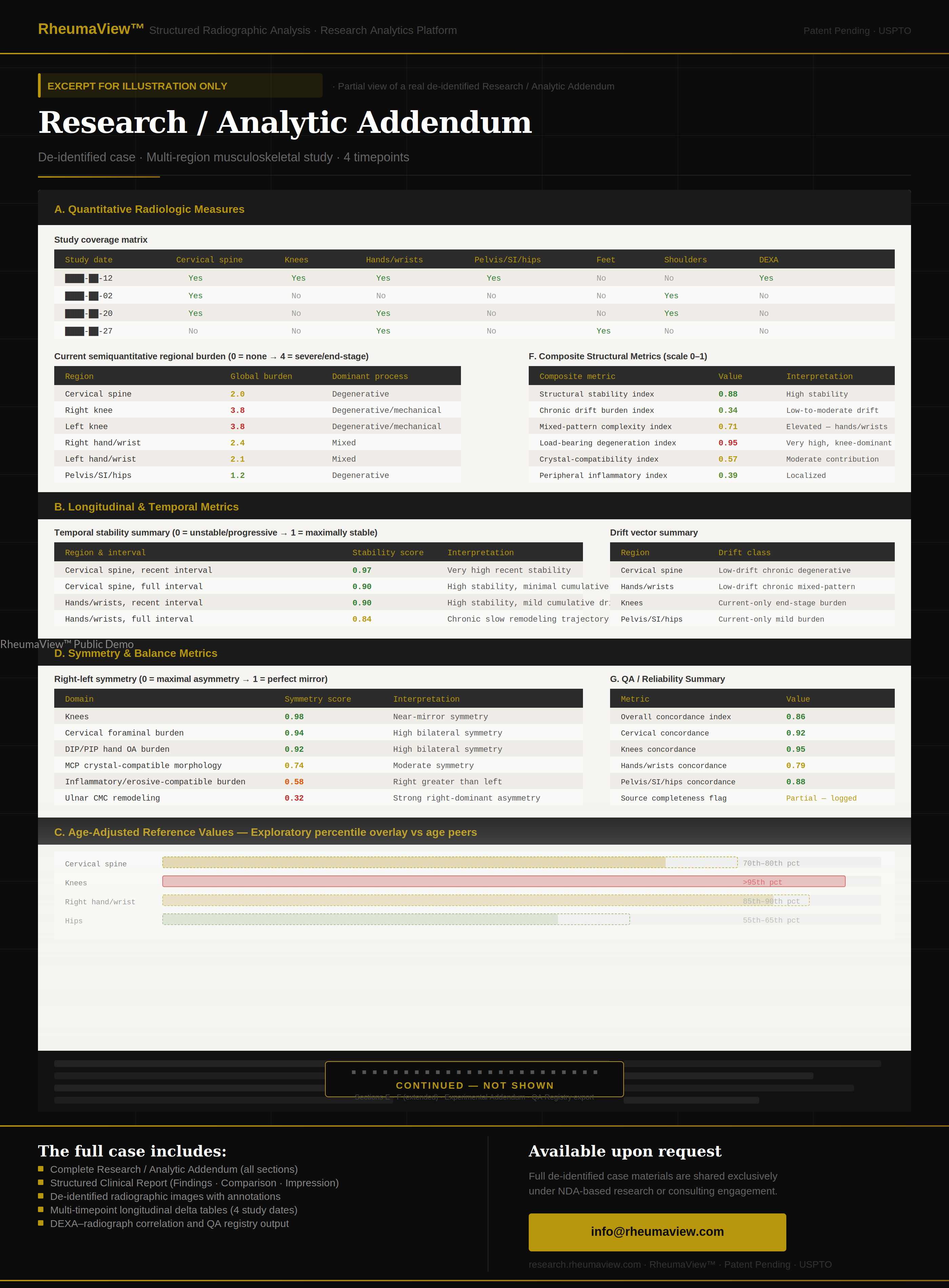
RheumaView™ addresses this at the architectural level. The platform operates through a validator-governed framework: structural descriptors, scoring vectors, and analytic outputs are not produced probabilistically — they are derived through a fixed, sequenced validation chain with full provenance tracing. The same input set yields reproducible, non-contradictory results across sessions, operators, and time elapsed between evaluations.
Three practical advantages
Methodological transparency
Every output carries traceable lineage — processing route, validation stage, and descriptor source are preserved and documentable. The answer to “how were findings derived?” is a defined, reproducible process, not a trained reader.
Inter-site consistency
Multi-institutional studies face the challenge of harmonizing data across readers and centers. Uniform analytical rules make cross-site structural data directly comparable — without post-hoc correction.
Reference framework
For work on imaging methodology — scoring-system development, endpoint validation, quantitative phenotyping — the platform provides a structured, reproducible reference layer to build against.
Why RheumaView™ fits research
Domains supported
Radiographic analysis spans single-specialty studies and cross-domain research designs:
Initiate a research consultation
Academic collaborations are structured through research service agreements with NDA-based access to methodology detail. Selected programs are invited to begin a scoped discussion.
Contact — Clinical & Research